 Basal cell carcinoma (BCC) is the most frequently diagnosed skin cancer. We often see it at our Hollywood dermatology practice. Understanding what basal cell carcinoma looks like and how it is treated can empower you to seek care early and prevent complications.
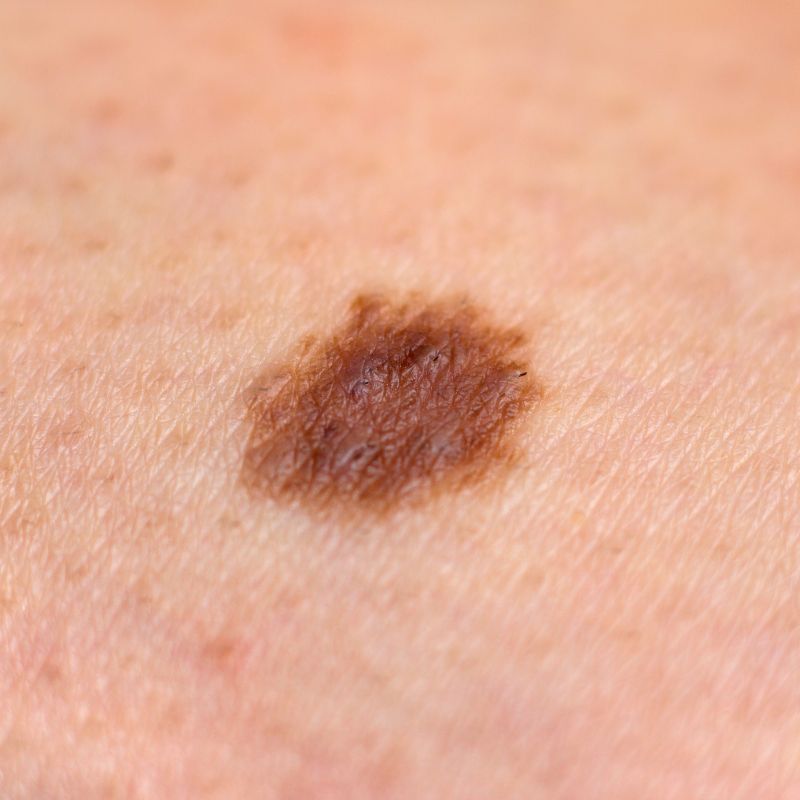
BCC typically presents as a small bump or growth on sun-exposed areas such as the face, neck, and arms. It may appear pearly or translucent, often with a pink or skin-colored hue, and sometimes features visible blood vessels or crusting. Some patients describe it as a persistent sore or a “pimple that doesn’t heal.” Unlike more aggressive cancers, basal cell carcinomas usually grow slowly over months or even years and rarely spread to other parts of the body.
Because basal cell carcinoma is so common and generally localized, early detection allows for straightforward treatment with excellent outcomes. If you notice a new growth or spot on your skin that bleeds, changes, or doesn’t heal, don’t delay seeing a dermatologist.
Diagnosing Basal Cell Carcinoma
When you visit our dermatology clinic with a suspicious lesion, we conduct a thorough skin examination. Using magnification tools, we can better visualize the lesion’s characteristics. To confirm the diagnosis, we usually perform a biopsy by numbing the area and removing a small piece of skin for lab analysis. Results are generally available within two weeks.
Treatment Options
If the biopsy confirms basal cell carcinoma, the treatment plan depends on the size, location, and depth of the lesion.
Scrape and Burn (Curettage and Electrodessication): This is a quick, in-office procedure where the cancerous tissue is scraped away, and the area cauterized to destroy any remaining cancer cells. It typically takes about 10 minutes and has a cure rate of over 90% when performed correctly. This method is particularly suitable for small, superficial basal cell carcinomas located on non-critical areas of the body.
Surgical Excision: For larger or more invasive tumors, we perform an excision in which the entire lesion is surgically removed, along with a margin of healthy skin. The wound is then closed with stitches. This method ensures complete removal because the tissue is sent to the path lab where they check for clear margins.
Mohs Micrographic Surgery: For lesions on the face or other sensitive areas, Mohs surgery is the gold standard. This technique involves removing the tumor layer by layer while examining the tissue under a microscope in real-time. The surgeon continues removing tissue only until no cancer cells remain. Mohs surgery spares as much healthy skin as possible, resulting in the smallest scar and preserving both appearance and function.
Basal Cell Carcinoma Follow-Up and Prevention
After treatment, regular follow-up visits every six months are crucial to monitor for any new skin cancers or recurrence. If you have risk factors such as a history of multiple sunburns, fair skin, or a prior skin cancer diagnosis, consistent skin checks can save your life.
Preventing basal cell carcinoma means adopting sun-safe habits. Daily use of a broad-spectrum sunscreen with at least SPF 30, wearing protective clothing, and avoiding peak sun hours are all effective strategies.
Remember, basal cell carcinoma rarely spreads, but can cause significant local damage if untreated. Early recognition and treatment ensure the best outcomes with minimal scarring. If you notice any suspicious bumps or sores on your skin, schedule an appointment as soon as possible.